
Laboratory Stand “Training Vehicle with Built-in Fault Simulator” (Copy)
July 30, 2025
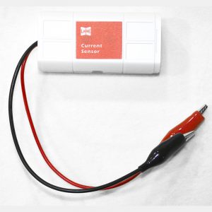
Wireless magnetic field sensor
August 1, 2025
Wireless ECG sensor
Range: 0~5V
Resolution: 0.1mV
Sampling Rate: 50-200Hz
I. Working Principle
The electrocardiogram, also known as a cardiac electrical activity diagram or EKG, represents the electrical signals generated by the heart. The electrocardiogram sensor is connected to your body to measure cardiac electrical activity, and the software displays this activity in graphical form.
II. Specifications
Range: 0~5V
Resolution: 0.1mV
Sampling Rate: 50-200Hz
III. Structure and Features
1. Equipped with a 1.8-inch color LCD screen.
2. Configured with five functional buttons for a simple and user-friendly interface.
3. Supports high-speed USB data transfer for rapid communication with data acquisition devices.
4. Enables wireless communication with experimental terminals.
5. Features a built-in high-capacity removable battery.
6. Includes a snap-on sensor interface for compatibility with standard sensors in combined experiments.
7. Provides fixed mounting points for integration with traditional equipment on iron stands.
8. The sensor is compatible with iOS, Android, and Windows systems.
IV. Typical Applications
The electrocardiogram sensor measures the voltage waveform (generated by heart contractions) of the heart.
Application Research:
– Measure electrocardiogram data while sitting, standing, before and after exercise, laughing, and before and after consuming caffeinated beverages.
Examples:
1. Monitor the electrocardiogram of Homo sapiens in a resting state.
2. Study the meanings of P, Q, R, S, and T waves.
3. Monitor the electrocardiogram after moderate exercise.
4. Investigate the electrocardiogram following mild stimulation.
V. Note
This product is intended solely for educational use and is not suitable for industrial, medical, research, or commercial applications.
VI. Usage Tips
1. Clean the area of skin where the electrocardiogram electrode will be attached.
2. Place the electrocardiogram electrode on the cleaned skin (use water, soapy water, or an alcohol swab for cleaning).
3. Position the electrodes as follows:
– Black ground electrode on the right forearm.
– White negative electrode on the right foot.
– Red positive electrode on the left forearm.
4. Metal or magnetic materials on the body may interfere with or distort the electrocardiogram signal.
5. Once the electrocardiogram leads are connected to the electrodes, the sensor is ready to acquire data.
(Note: The baseline of the electrocardiogram waveform may shift during sensor data collection due to movement of large muscle groups. For optimal results, remain still while collecting data.)

Reviews
There are no reviews yet.